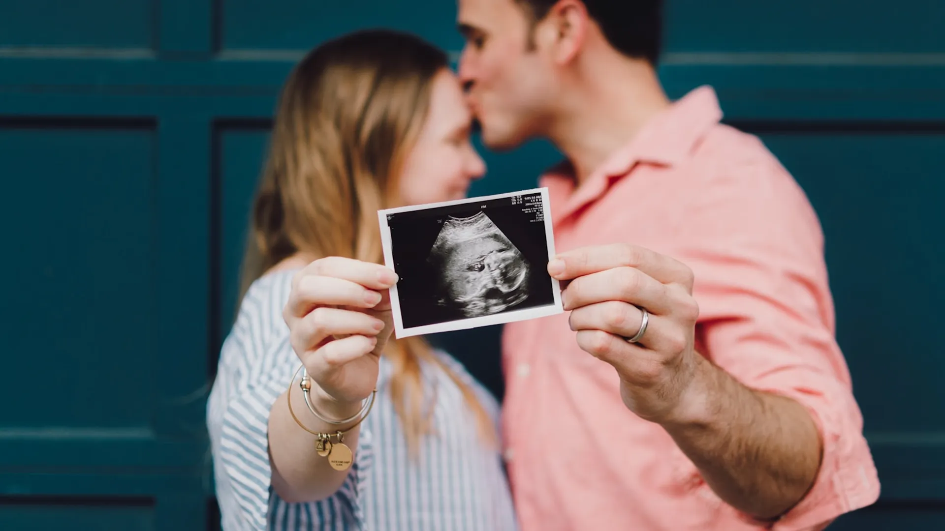
Prenatal diagnosis of cleft lip and palate makes it possible to identify, before birth, a difference in the development of the face, and to begin preparing appropriately for the care of the newborn.
In most cases, the first suspicion arises during the anomaly scan (20-week scan) in the second trimester, usually at around 20 weeks of pregnancy. However, not all clefts are identified equally easily. What an ultrasound can show depends on the type and extent of the cleft, the position of the baby, the gestational age and the conditions of the examination, as well as the experience of the professional who performs and interprets the scan. [1–5]
For this reason, prenatal diagnosis should not be limited to answering whether or not a cleft is present. The aim is to characterise which structures are affected, assess whether other associated anomalies exist, and give the family the information they need to plan the care of the newborn. [1,4]
When can cleft lip and palate be detected during pregnancy?
The anomaly scan in the second trimester, usually carried out at around 20 weeks, is one of the main opportunities to detect cleft lip and palate.
During this examination, the obstetric team makes a systematic assessment of the baby’s anatomy. The assessment of the face includes the facial profile and the upper lip, among other structures.
A break in the anatomical continuity of the lip, seen on ultrasound, can raise suspicion of the diagnosis. However, the ability to detect a cleft varies considerably depending on its type.
A population-based study in Denmark, which included 994 cases of orofacial clefts, found a prenatal detection rate of 71.7% for cleft lip with or without cleft palate. When the different types were analysed separately, detection was 76.6% for unilateral cleft lip and palate and 82.3% for bilateral. By contrast, only 3.3% of isolated cleft palates were identified before birth. [3]
These figures show that not all cases of cleft lip and palate can be detected during pregnancy, especially isolated cleft palate.
Although some clefts can be identified before 20 weeks using specialised ultrasound protocols, the second-trimester anomaly scan remains a key moment for the prenatal assessment of facial anatomy. [1,3,5]
How is cleft lip and palate detected before birth?
Prenatal diagnosis usually begins with the obstetric team, and especially with the professional specialised in fetal ultrasound who performs and interprets the scan.
The diagnostic power of ultrasound does not depend only on the device used. The type and extent of the cleft, the gestational age, the position of the baby and the anatomical conditions that allow images to be obtained also play a part. The experience of the operator is also very important. [1,3,5]
When a cleft is suspected and cannot be adequately characterised with conventional ultrasound, a specialised fetal ultrasound assessment may be indicated.
This assessment allows the facial anatomy to be studied in greater detail, so that the suspected diagnosis of cleft lip and palate can be confirmed. Advanced ultrasound also makes it possible to assess the rest of the baby’s anatomy to determine whether other associated anomalies are present. [1,4]
Can a 2D anomaly scan confirm whether the palate is affected?
Yes, in part. Two-dimensional (2D) ultrasound is the technique routinely used to assess the baby, and it can identify most clefts that affect the lip.
In a cleft lip, the break in the continuity of the upper lip can be visible on ultrasound. However, determining whether the palate is also affected is considerably more complex.
The baby’s palate requires specific scanning planes, and its visualisation can be limited by the position of the baby and by bony structures that make certain images difficult to obtain. For this reason, ultrasound is very limited in its ability to detect an isolated cleft palate. [2,3,5]
As a result, identifying a cleft of the lip does not always make it possible to determine with certainty, using conventional ultrasound, whether the palate is also affected.
When is a 3D or 4D ultrasound indicated?
When a cleft lip and palate is suspected, an advanced fetal ultrasound that includes three-dimensional (3D) reconstructions can provide additional information.
3D ultrasound makes it possible to reconstruct the facial anatomy from different planes and can help characterise the cleft and structures such as the lip and the alveolar ridge. In certain circumstances it can also provide complementary information for assessing the palate. [1,5]
The available evidence indicates that adding 3D techniques can improve the characterisation of some clefts compared with conventional ultrasound, but the results depend on the quality of the images obtained, the experience of the operator and the type of cleft. [2,5]
For this reason, 3D/4D ultrasound should not be considered mandatory for every pregnancy, nor a substitute for conventional obstetric ultrasound. Its main value is to complement a specialised assessment when a cleft is suspected or when the facial anatomy needs to be defined more precisely.
What happens after a cleft lip and palate is detected on a pregnancy scan?
Once a cleft has been identified, the next step is to assess the rest of the baby’s anatomy to check that there are no additional anatomical anomalies that would require further genetic testing. [1,4]
In more than 90% of cleft lip and palate cases detected by ultrasound, the cleft is an isolated anomaly, with no other identifiable anatomical differences.
Is genetic testing necessary?
Not every family in which a cleft lip and palate is identified necessarily needs invasive genetic testing.
The decision to carry out amniocentesis or chorionic villus sampling should be individualised according to the type of cleft, the findings of the fetal ultrasound, the presence of other anatomical anomalies and the family history. [1,4]
For this reason, a diagnosis of cleft lip and palate does not automatically mean that amniocentesis or chorionic villus sampling is necessary. The decision should form part of an individualised prenatal assessment, usually involving maternal-fetal medicine and, where indicated, clinical genetics. [1,4]
In summary
The anomaly scan at around 20 weeks is one of the main opportunities to detect cleft lip and palate during pregnancy.
2D ultrasound can identify many clefts of the lip, but involvement of the palate is harder to assess. This difficulty is especially marked in isolated cleft palate, whose prenatal detection remains limited in population screening. [2,3]
When a cleft is suspected, a specialised fetal ultrasound, complemented when indicated by 3D/4D reconstructions, can provide additional information and help characterise the facial anatomy better. In high-risk settings and with targeted examinations, the accuracy for identifying palate involvement can be considerably higher than that seen in routine screening. [2,5]
After a cleft is detected, the aim should be broader than confirming its presence: it must be characterised, the rest of the baby’s anatomy assessed, and any associated findings identified that could change the counselling and the need for genetic testing. [1,4]
In short, prenatal diagnosis of cleft lip and palate is not simply about “seeing a cleft” on an ultrasound. It is about understanding which structures are affected, assessing the baby’s clinical context, and giving the family rigorous, understandable information so they can begin planning the care of their newborn.